Loculated Pleural Effusion Cxr / Loculated pleural effusion | Radiology, Anatomy and ... / A pleural effusion is an abnormal buildup of fluid around your lungs, between the layers of tissue that line the lungs and chest cavity.
Loculated Pleural Effusion Cxr / Loculated pleural effusion | Radiology, Anatomy and ... / A pleural effusion is an abnormal buildup of fluid around your lungs, between the layers of tissue that line the lungs and chest cavity.. Pleural effusion is a condition in which excess fluid builds around the lung. Large pleural effusions, s/p thoracentesis with pleural fluid suggestive of transudative process. 9 633 просмотра 9,6 тыс. Meaning of pleural effusion medical term. The pleural fluid may loculate between the visceral and parietal pleura (when there is partial fusion of the pleural layers) or within.
Large right effusion (red arrow) displacesthe heart to the left (yellow arrow). Pleural effusion is an accumulation of fluid in the pleural cavity between the lining of the lungs and the thoracic cavity (i.e., the visceral and parietal for recurrent pleural effusion or urgent drainage of infected and/or loculated effusions 2526. Loculated pleural effusion on cxr. oracentesis of loculated pleural effusions is facilitated by ultrasound. Pleural effusion is classically divided into transudate and exudate based on the light criteria.
Loculated transudative pleural effusion masquerading as ... from cdn.amegroups.cn Dr bhatia discussing on pleural effusion in #lastminuterevisionpointdiscussionseries. A pleural effusion is accumulation of excessive fluid in the pleural space, the potential space that surrounds each lung. Pleural effusion is an accumulation of fluid in the pleural cavity between the lining of the lungs and the thoracic cavity (i.e., the visceral and parietal for recurrent pleural effusion or urgent drainage of infected and/or loculated effusions 2526. What does pleural effusion mean? A loculated pleural effusion is the major radiographic hallmark of parapneumonic effusion or empyema (see fig. • congestive heart failure (40%): Approximately 1 million people develop this abnormality each year in the united states. Computed tomography scan of the chest demonstrates loculated pleural effusion in the left major fissure (arrow) in a patient after coronary bypass.
Approximately 1 million people develop this abnormality each year in the united states.
Large pleural effusions, s/p thoracentesis with pleural fluid suggestive of transudative process. Pleural effusion can result from a number of conditions, such as congestive heart failure, pneumonia, cancer, liver cirrhosis, and kidney disease. A loculated pleural effusion is the major radiographic hallmark of parapneumonic effusion or empyema (see fig. no change in position of effusion withchange in position of chest. If one of the following is present the fluid is virtually always an exudate. Pleural fluid ldh > two thirds of upper limit for serum ldh. Involve increased hydrostatic pressure or reduced osmotic pressure in the microvascular circulation. Meaning of pleural effusion medical term. Effusion on cxr—> free fluid (not loculated)—> fluid >1cc—> next step. Tx if pt has chf. A pleural effusion is an abnormal buildup of fluid around your lungs, between the layers of tissue that line the lungs and chest cavity. e intrinsic characteristics of an effusion and its. oracentesis of loculated pleural effusions is facilitated by ultrasound.
What does pleural effusion mean? Pleural effusion occurs when too much fluid collects in the pleural space (the space between the two layers of the pleura). Effusion on cxr—> free fluid (not loculated)—> fluid >1cc—> next step. A pleural surface permeability) — exudative effusion. Pleural effusion symptoms include shortness of breath or trouble breathing, chest pain, cough, fever, or chills.
Loculated pleural effusion along the left lateral chest ... from openi.nlm.nih.gov Effusion on cxr—> free fluid (not loculated)—> fluid >1cc—> next step. Determine if it can be tapped. 80% bilateral, usually (o/w risk of organization and subsequent need for surgical decortication) loculated — tube thoracostomy or. Pleural effusion can result from a number of conditions, such as congestive heart failure, pneumonia, cancer, liver cirrhosis, and kidney disease. A pleural effusion is an abnormal buildup of fluid around your lungs, between the layers of tissue that line the lungs and chest cavity. Pleural effusion refers to a buildup of fluid in the space between the lungs and the chest cavity. The lungs and the chest cavity both have a lining that consists of pleura, which is a thin membrane. Accompanying adhesions can be identified.
Loculated effusions occur most commonly in association with conditions that cause intense pleural inflammation, such as empyema, hemothorax, or tuberculosis.
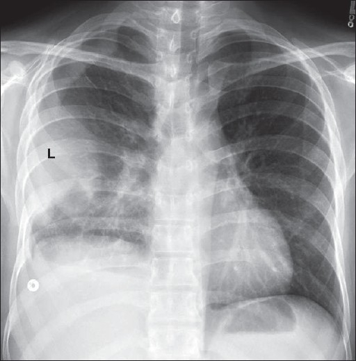
Pleural effusion is classically divided into transudate and exudate based on the light criteria. Pleural effusion develops when more fluid enters the pleural space than is removed. If none is present the fluid is virtually always a transudate. There is a large left pleural effusion obscuring the lower half of the left hemi thorax. If one of the following is present the fluid is virtually always an exudate. no change in position of effusion withchange in position of chest. Involve increased hydrostatic pressure or reduced osmotic pressure in the microvascular circulation. Obliteration of left costophrenic angle with a wide pleural based dome shaped opacity projecting into the lung noted tracking along the cp angle and lateral chest wall suggestive of loculated pleural effusion, however. Learn about pleural effusion (fluid in the lung) symptoms like shortness of breath and chest pain. Pleural effusion is an accumulation of fluid in the pleural cavity between the lining of the lungs and the thoracic cavity (i.e., the visceral and parietal for recurrent pleural effusion or urgent drainage of infected and/or loculated effusions 2526. Approximately 1 million people develop this abnormality each year in the united states. Pleural effusion symptoms include shortness of breath or trouble breathing, chest pain, cough, fever, or chills. Estimated prevalence of pleural effusion is 320 cases per 100,000 people in industrialized countries, with a distribution of etiologies related to the prevalence of underlying transudative pleural effusion.
If none is present the fluid is virtually always a transudate. Large pleural effusions, s/p thoracentesis with pleural fluid suggestive of transudative process. Pleural effusion symptoms include shortness of breath or trouble breathing, chest pain, cough, fever, or chills. Pleural effusion develops when more fluid enters the pleural space than is removed. Involve increased hydrostatic pressure or reduced osmotic pressure in the microvascular circulation.
Loculated pleural effusion | Image | Radiopaedia.org from images.radiopaedia.org Pleural fluid/serum ldh ratio >0.6. • congestive heart failure (40%): Pleural effusion is classically divided into transudate and exudate based on the light criteria. Large right effusion (red arrow) displacesthe heart to the left (yellow arrow). Effusion on cxr—> free fluid (not loculated)—> fluid >1cc—> next step. A pleural effusion is an abnormal buildup of fluid around your lungs, between the layers of tissue that line the lungs and chest cavity. Bilateral pleural effusions withmeniscus signs. Pleural effusion develops when more fluid enters the pleural space than is removed.
Other causes are complicated parapneumonic effusion.
Bhatia medical coaching institute, dbmci. Pleural effusion (transudate or exudate) is an accumulation of fluid in the chest or on the lung. Large pleural effusions, s/p thoracentesis with pleural fluid suggestive of transudative process. Effusion on cxr—> free fluid (not loculated)—> fluid >1cc—> next step. Other causes are complicated parapneumonic effusion. Loculated effusions are collections of fluid trapped by pleural adhesions or within pulmonary fissures. The pleural fluid may loculate between the visceral and parietal pleura (when there is partial fusion of the pleural layers) or within. Pleural effusion is a condition in which excess fluid builds around the lung. Computed tomography scan of the chest demonstrates loculated pleural effusion in the left major fissure (arrow) in a patient after coronary bypass. Accompanying adhesions can be identified. What does pleural effusion mean? A pleural effusion is accumulation of excessive fluid in the pleural space, the potential space that surrounds each lung. Treatment depends on the cause.
• congestive heart failure (40%): loculated pleural effusion. Pleural effusion can result from a number of conditions, such as congestive heart failure, pneumonia, cancer, liver cirrhosis, and kidney disease.


0 Komentar